CODAIT / Deep Histopath
Projects that are alternatives of or similar to Deep Histopath
Predicting Breast Cancer Proliferation Scores with TensorFlow, Keras, and Apache Spark
Note: This project is still a work in progress. There is also an experimental branch with additional files and experiments.
Overview
The Tumor Proliferation Assessment Challenge 2016 (TUPAC16) is a "Grand Challenge" that was created for the 2016 Medical Image Computing and Computer Assisted Intervention (MICCAI 2016) conference. In this challenge, the goal is to develop state-of-the-art algorithms for automatic prediction of tumor proliferation scores from whole-slide histopathology images of breast tumors.
Background
Breast cancer is the leading cause of cancerous death in women in less-developed countries, and is the second leading cause of cancerous deaths in developed countries, accounting for 29% of all cancers in women within the U.S. [1]. Survival rates increase as early detection increases, giving incentive for pathologists and the medical world at large to develop improved methods for even earlier detection [2]. There are many forms of breast cancer including Ductal Carcinoma in Situ (DCIS), Invasive Ductal Carcinoma (IDC), Tubular Carcinoma of the Breast, Medullary Carcinoma of the Breast, Invasive Lobular Carcinoma, Inflammatory Breast Cancer and several others [3]. Within all of these forms of breast cancer, the rate in which breast cancer cells grow (proliferation), is a strong indicator of a patient’s prognosis. Although there are many means of determining the presence of breast cancer, tumor proliferation speed has been proven to help pathologists determine the best treatment for the patient. The most common technique for determining the proliferation speed is through mitotic count (mitotic index) estimates, in which a pathologist counts the dividing cell nuclei in hematoxylin and eosin (H&E) stained slide preparations to determine the number of mitotic bodies. Given this, the pathologist produces a proliferation score of either 1, 2, or 3, ranging from better to worse prognosis [4]. Unfortunately, this approach is known to have reproducibility problems due to the variability in counting, as well as the difficulty in distinguishing between different grades.
References:
[1] http://emedicine.medscape.com/article/1947145-overview#a3
[2] http://emedicine.medscape.com/article/1947145-overview#a7
[3] http://emedicine.medscape.com/article/1954658-overview
[4] http://emedicine.medscape.com/article/1947145-workup#c12
Goal & Approach
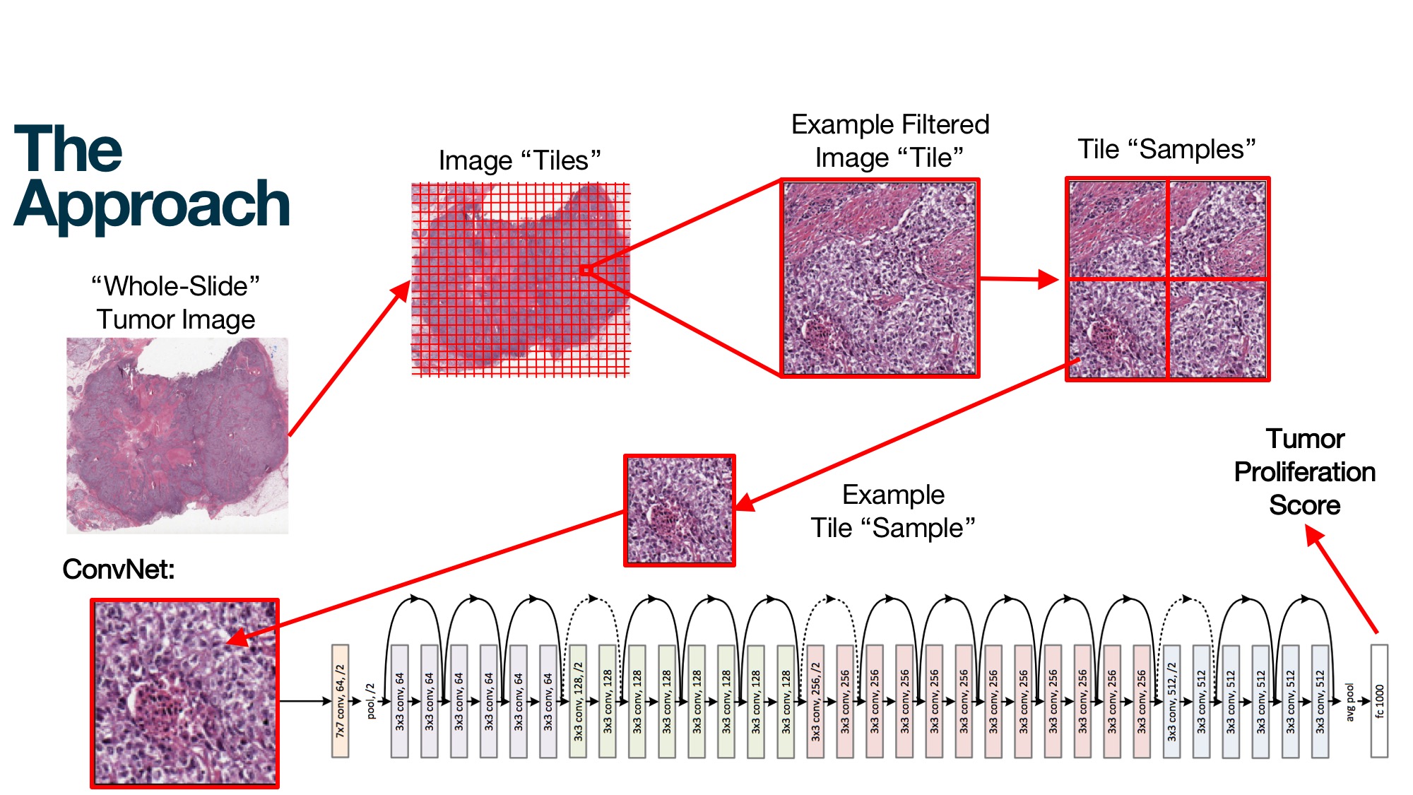
In an effort to automate the process of classification, this project aims to develop a large-scale deep learning approach for predicting tumor scores directly from the pixels of whole-slide histopathology images (WSI). Our proposed approach is based on a recent research paper from Stanford [1]. Starting with 500 extremely high-resolution tumor slide images [2] with accompanying score labels, we aim to make use of Apache Spark in a preprocessing step to cut and filter the images into smaller square samples, generating 4.7 million samples for a total of ~7TB of data [3]. We then utilize TensorFlow and Keras to train a deep convolutional neural network on these samples, making use of transfer learning by fine-tuning a modified ResNet50 model [4]. Our model takes as input the pixel values of the individual samples, and is trained to predict the correct tumor score classification for each one. We also explore an alternative approach of first training a mitosis detection model [5] on an auxiliary mitosis dataset, and then applying it to the WSIs, based on an approach from Paeng et al. [6]. Ultimately, we aim to develop a model that is sufficiently stronger than existing approaches for the task of breast cancer tumor proliferation score classification.
References:
[1] https://web.stanford.edu/group/rubinlab/pubs/2243353.pdf
[2] http://tupac.tue-image.nl/node/3
[3] preprocess.py, breastcancer/preprocessing.py
[4] MachineLearning-Keras-ResNet50.ipynb
[5] preprocess_mitoses.py, train_mitoses.py
[6] https://arxiv.org/abs/1612.07180
Setup (All nodes unless other specified):
-
System Packages:
openslide
-
Python packages:
- Basics
pip3 install -U matplotlib numpy pandas scipy jupyter ipython scikit-learn scikit-image openslide-python
- TensorFlow (only on driver):
-
pip3 install tensorflow-gpu(orpip3 install tensorflowfor CPU-only)
-
- Keras (bleeding-edge; only on driver):
pip3 install git+https://github.com/fchollet/keras.git
- Basics
-
Spark 2.x (ideally bleeding-edge)
-
Add the following to the
datafolder (same location on all nodes):-
training_image_datafolder with the training slides. -
testing_image_datafolder with the testing slides. -
training_ground_truth.csvfile containing the tumor & molecular scores for each slide. -
mitosesfolder with the following from the mitosis detection auxiliary dataset:-
mitoses_test_image_datafolder with the folders of testing images -
mitoses_train_image_datafolder with the folders of training images -
mitoses_train_ground_truthfolder with the folders of training csv files
-
-
-
Layout:
- MachineLearning-Keras-ResNet50.ipynb - breastcancer/ - preprocessing.py - visualization.py - ... - data/ - mitoses - mitoses_test_image_data - 01 - 01.tif - 02 - 01.tif ... - mitoses_train_ground_truth - 01 - 01.csv - 02.csv ... - 02 - 01.csv - 02.csv ... ... - mitoses_train_image_data - 01 - 01.tif - 02.tif ... - 02 - 01.tif - 02.tif ... ... - training_ground_truth.csv - training_image_data - TUPAC-TR-001.svs - TUPAC-TR-002.svs - ... - testing_image_data - TUPAC-TE-001.svs - TUPAC-TE-002.svs - ... - preprocess.py - preprocess_mitoses.py - train_mitoses.py -
Adjust the Spark settings in
$SPARK_HOME/conf/spark-defaults.confusing the following examples, depending on the job being executed:-
All jobs:
# Use most of the driver memory. spark.driver.memory 70g # Remove the max result size constraint. spark.driver.maxResultSize 0 # Increase the message size. spark.rpc.message.maxSize 128 # Extend the network timeout threshold. spark.network.timeout 1000s # Setup some extra Java options for performance. spark.driver.extraJavaOptions -server -Xmn12G spark.executor.extraJavaOptions -server -Xmn12G # Setup local directories on separate disks for intermediate read/write performance, if running # on Spark Standalone clusters. spark.local.dirs /disk2/local,/disk3/local,/disk4/local,/disk5/local,/disk6/local,/disk7/local,/disk8/local,/disk9/local,/disk10/local,/disk11/local,/disk12/local -
Preprocessing:
# Save 1/2 executor memory for Python processes
spark.executor.memory 50g
-
-
To execute the WSI preprocessing script, use
spark-submitas follows (could also use Yarn in client mode with--master yarn --deploy-mode client):PYSPARK_PYTHON=python3 spark-submit --master spark://MASTER_URL:7077 preprocess.py -
To execute the mitoses preprocessing script, use the following:
python3 preprocess_mitoses.py --help -
To execute the mitoses training script, use the following:
python3 training_mitoses.py --help -
To use the Jupyter notebooks, start up Jupyter like normal with
jupyter notebookand run the desired notebook.
Create a Histopath slide “lab” to view the slides (just driver):
git clone https://github.com/openslide/openslide-python.git- Host locally:
python3 path/to/openslide-python/examples/deepzoom/deepzoom_multiserver.py -Q 100 path/to/data/
- Host on server:
python3 path/to/openslide-python/examples/deepzoom/deepzoom_multiserver.py -Q 100 -l HOSTING_URL_HERE path/to/data/- Open local browser to
HOSTING_URL_HERE:5000.

