threeBrain - HTML, WebGL based 3D Viewer
![]()


Key Features:
- Uses modern browsers, easy to embed and share
- Displays MRI, surfaces, and electrodes in the same canvas
- Maps multiple subjects on template brains using
AFNI/SUMA(standard 141) orMNI-305locations - Electrode localization in 3 approaches
- Volume rendering and surface/electrode animation
- Integration with interactive
R-shinyframework
News | reference page | keyboard shortcuts
System Requirement
- Web Browsers: the viewer uses
WegGL2to render in browsers. Please check this list to see compatible browsers. As of 2020, Chrome, Firefox, and Edge (not IE) have full supports.- For Safari users, please enable this feature by going to
Safari>Preferences, clickAdvanced, then selectShow Develop menu in menu bar; then clickDevelopin the menu bar, go toExperimental Features>WebGL 2.0. This only needs to be done once.
- For Safari users, please enable this feature by going to
A. Installation
RandRStudio Desktop (Free Version)- Open
RStudio, enter from its console:
install.packages("threeBrain")If you want to install dev version from Github, then use:
install.packages("remotes")
remotes::install_github("dipterix/threeBrain")- (Optional) Setups: after installation, in
RStudioconsole, type the following command
threeBrain::brain_setup()and follow the instructions.
B. Basic Brain Viewer
Once finishing setting up of threeBrain, there will be a template subject N27 (Collin's 27) created locally. The location is platform-related. You can find it by running the following command:
library(threeBrain)

default_template_directory()
#> [1] "/Users/dipterix/Library/Application Support/
#> org.R-project.R/R/threeBrain/templates"
N27 template folder resides inside of this directory.
Let's view this subject using the freesurfer_brain2 function.
- Import subject
library(threeBrain)

n27_path <- file.path(default_template_directory(), "N27")

x <- freesurfer_brain2( fs_subject_folder = n27_path,
subject_name = 'N27', surface_types = 'pial')- Visualize
plot(x) # alternatively, you can use `n27$plot()`C. Subject Setup
The sample subject (N27) is a sample generated by FreeSurfer (download). If you have any subjects processed by FreeSurfer, use function freesurfer_brain2 to visualize.
The AFNI/SUMA standard 141 brain is also supported. Please use terminal command @SUMA_Make_Spec_FS -NIFTI -sid [subID] to generate 141 brain. (Click here for some hints)
D. Add/Render Electrodes
If you have electrode file, you can import it before calling plot function. Please make sure it's in csv format.
x$set_electrodes(electrodes = "[PATH to ELECTRODE FILE]")Here is an example of electrode csv file. Only the first five columns (case-sensitive) are mandatory: Electrode (integer), Coord_x, Coord_y, Coord_z, and Label (character). Coord_* is tkRAS location from FreeSurfer coordinates.
| Electrode| Coord_x| Coord_y| Coord_z|Label  | MNI305_x|  MNI305_y|  MNI305_z|SurfaceElectrode |SurfaceType | Radius| VertexNumber|Hemisphere |
|---------:|-------:|-------:|-------:|:------|--------:|---------:|---------:|:----------------|:-----------|------:|------------:|:----------|
|         1|    29.0|    -7.8|   -34.6|RMHCH1 | 30.46817| -17.98119| -23.40022|FALSE            |pial        |      2|           -1|left       |
|         2|    33.8|    -8.0|   -34.2|RMHCH2 | 35.57109| -17.76624| -22.80131|FALSE            |pial        |      2|           -1|left       |
|         3|    38.0|    -7.5|   -33.5|RMHCH3 | 39.97102| -16.81249| -22.17986|FALSE            |white       |      2|           -1|right      |
|         4|    42.6|    -6.8|   -33.0|RMHCH4 | 44.79092| -15.73442| -21.82591|FALSE            |smoothwm    |      2|           -1|right      |
|         5|    47.0|    -6.8|   -32.6|RMHCH5 | 49.45370| -15.35431| -21.31272|FALSE            |pial        |      2|           -1|right      |
|         ...
To assign values to electrodes, run
x$set_electrode_values(electrodes = "[PATH to ELECTRODE VALUE FILE]")The electrode value file is also a csv like:
| Electrode| Subject| Project|    Time| ValueName| ValueName2|  ...|
|---------:|-------:|-------:|-------:|:---------|----------:|-----|
|         1|     N27|    Demo|       0|A         |   30.46817|  ...|
|         2|     N27|    Demo|       0|B         |   35.57109|  ...|
|         3|     N27|    Demo|       0|C         |   39.97102|  ...|
|         4|     N27|    Demo|       0|D         |   44.79092|  ...|
|         5|     N27|    Demo|       0|A         |   49.45370|  ...|
|         ...
Project and Time are optional. However, if you are also using rave, please make sure Project exists. If you want to show animation, Time is necessary and must be numeric. ValueName? can be any characters containing letters (A-Z, a-z), letters (0-9) and underscore (_).
E. Merge Subjects and Template mapping
If you have your own subjects with FreeSurfer output, for example, I have two subjects YAB and YCQ. To merge these two subjects and show them on N27 template,
library(threeBrain)

# yab = ... (see section B for import a single subject)
# ycq = ...
template_n27 = merge_brain(yab, ycq, template_subject = 'N27')

plot( template_n27 )
The viewer will be in N27 template, and electrodes of these two subjects can be mapped via MNI305 (for surface and stereo EEG) or std.141 (for surface-only).
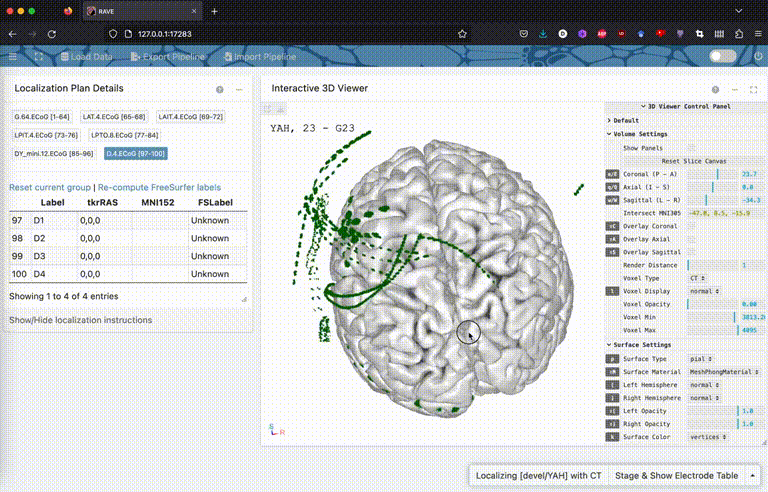
F. Electrode Localization
(Do NOT use this feature for clinical purposes!)
As of version 0.2.1, threeBrain supports electrode localization. You can:
- Use template brain to generate electrode table, when you don't have subject-level MRI
- Use subject MRI to generate when CT scans are unavailable
- Use CT co-registered to MRI
Use template brain to generate electrode table
Create a blank template and localize:
library(threeBrain)
template <- merge_brain()
template$localize()
Use subject MRI to generate when CT scans are unavailable
If you have MRI but don't have CT scans, it is possible to use MRI slices. Make sure to change fs_subject_folder accordingly:
x <- freesurfer_brain2(
  fs_subject_folder = file.path(default_template_directory(), "N27"),
  subject_name = 'N27', surface_types = 'pial')

x$localize()
Use CT co-registered to MRI
If you have CT images, please co-register with MRI first. This requires dcm2nii (link) or dcm2niix (link), and FLIRT package (link).
Step 1: Merge all DICOM images to Nifti format:
Open your terminal, change directories to where CT images are, and run
dcm2niix [folder with DICOM images]
Do the same to T1 MR images too.
Step 2: Copy the two .nii files generated in the previous step to a same folder, rename them to be ct.nii and t1.nii
flirt -in ct.nii -ref t1.nii -out ct_in_t1.nii -omat ct2t1.mat -interp trilinear -cost mutualinfo -dof 6 -searchcost mutualinfo -searchrx -180 180 -searchry -180 180 -searchrz -180 180
There will be a ct_in_t1.nii file generated.
Step 3: Localize
Open RStudio, type and change the file path to ct_in_t1.nii just created
# Import 3D brain
# x <- freesurfer_brain2( ... )
x$localize( "[path to ct_in_t1.nii]" )
Citation
To cite threeBrain in publications use:
Magnotti, J. F., Wang, Z., & Beauchamp, M. S. (2020). RAVE: Comprehensive open-source software for reproducible analysis and visualization of intracranial EEG data. NeuroImage, 223, 117341.
A BibTeX entry for LaTeX users:
  @Article{,
    title = {{RAVE}: Comprehensive open-source software for reproducible analysis and visualization of intracranial EEG data},
    author = {John F. Magnotti and Zhengjia Wang and Michael S. Beauchamp},
    journal = {NeuroImage},
    year = {2020},
    volume = {223},
    doi = {10.1016/j.neuroimage.2020.117341},
    pages = {117341},
  }